MedFoxes

Search
Close this search box.

Structural Heart

Cardiac Structures in Intracardiac Echocardiography

Intracardiac echocardiography (ICE) is a cutting-edge diagnostic tool that offers an unparalleled view of the heart’s anatomy and function during electrophysiological procedures. This technique allows physicians to visualize the heart from within, providing detailed images of the cardiac structures that are crucial for successful electrophysiology studies. In this article, we will explore the role of […]

Cardiac Structures in Intracardiac Echocardiography Read More »

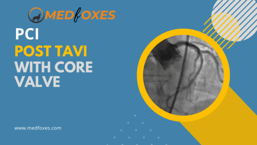
PCI Post TAVI with Core Valve

Unplanned PCI post TAVI is uncommon, and the incidence decreases over time. In the first two years following TAVI, acute coronary syndrome is the primary rationale for PCI; thereafter, chronic coronary syndromes are more common. Balloon-expandable and self-expandable bioprostheses appear to have no differences in the frequency or success of unplanned PCIs after TAVR. Hardware’s

PCI Post TAVI with Core Valve Read More »

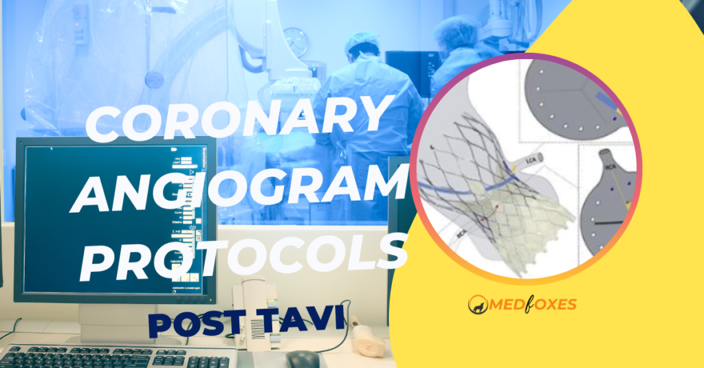
Coronary Angiogram Protocols Post TAVI With Core Valve

Following TAVI, coronary interventions can be technically challenging. The selective cannulation of the coronary ostia, which is dependent on patient anatomy, valve type and design, and implantation parameters including implantation depth and the orientation of the commissural tabs with respect to the ostia, is the most difficult part of the surgery. The following, we summarise

Coronary Angiogram Protocols Post TAVI With Core Valve Read More »

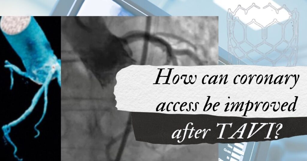
How can coronary access be improved after TAVI?

The prevalence of coronary artery disease in patients with severe aortic stenosis undergoing TAVI ranges from 40% to 75%. According to single-center studies, between 3.5% and 5.7% of TAVI patients required PCI after TAVI. “As the indication for TAVR expands to lower risk patients with a better long-term prognosis, the need for repeat coronary angiography and

How can coronary access be improved after TAVI? Read More »

Intra-OP Imaging During TAVI

TAVI, just like any structural heart disease intervention, relies on effective imaging earlier, throughout, and after the procedure. Imaging’s role in percutaneous valve replacement is comparable to that of appropriate exposure and visualisation in open surgical valve replacement. Visualisation improves the speed, safety, and predictability of the procedure. In addition, imaging throughout the procedure is

Intra-OP Imaging During TAVI Read More »

Magnetic Resonance Imaging on TAVI

Cardiac Magnetic Resonance Imaging is a three-dimensional technology that uses magnetic fields and radio waves to acquire detailed pictures of a patient’s heart.Unlike certain other imaging techniques, it doesn’t use ionising radiation, so patients don’t need to be exposed to harmful X-rays or radioactive substances. In the assessment of aortic valve disease, cardiac MRI yields

Magnetic Resonance Imaging on TAVI Read More »

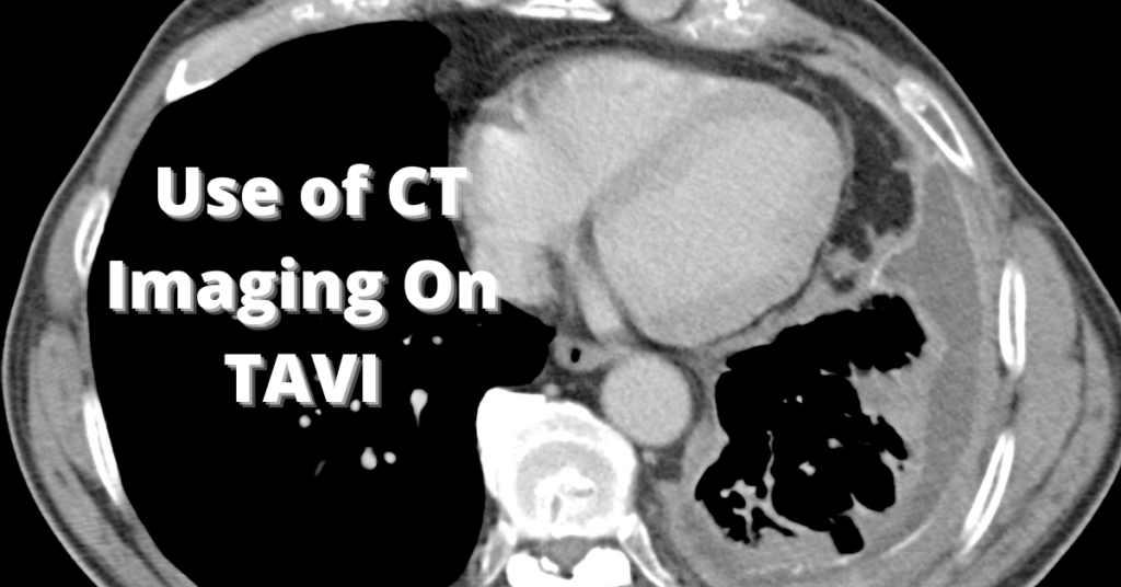
Multislice CT Imaging on Transcatheter Aortic Valve Implantation

Multislice computed tomography (MultiSlice CT) is the most comprehensive and non-invasive imaging modality to evaluate candidates for transcatheter aortic valve implantation. It can help confirm the severity of aortic stenosis and assess procedural feasibility, as well as device sizing. Evaluation of the Peripheral Arteries and Aorta Procedures to implant transcatheter aortic valves, particularly when done

Multislice CT Imaging on Transcatheter Aortic Valve Implantation Read More »

How Does Imaging Affect Transcatheter Aortic Valve Implantation?​

In the past few years, transcatheter aortic valve implantation has been one of the main breakthroughs in the treatment of patients with symptomatic, severe aortic stenosis who are at high risk for surgery. In patients with either self-expandable or balloon-expandable valvular prostheses, transcatheter aortic valve implantation has shown to be technically feasible and has provided

How Does Imaging Affect Transcatheter Aortic Valve Implantation?​ Read More »