

ECG Foundation Crash Course – Master the Basics Fast
FREE
ECG Foundation Crash Course – Master the Basics Fast Read More »

499$ – 6Months Program,
699$ – 9Months Program,
1200$ one on one.
Cardiac Electrophysiology Masterclass Read More »

399$ – 6Months Program,
499$ – 9Months Program,
999$ one on one.
Comprehensive Cardiovascular Technology Masterclass Read More »
Transeptal puncture (TSP) is a crucial step in numerous electrophysiology procedures, including atrial fibrillation ablation, left atrial (LA) and left ventricular (LV) mapping and ablation, and various interventional procedures like implantation of left atrial appendage (LAA) occlusion devices or mitral valve modifying clips, with transseptal puncture being the most commonly employed method for gaining access
Improving Transeptal Puncture Accuracy and Safety: The Role of ICE Imaging Read More »

Intracardiac echocardiography (ICE) is a cutting-edge diagnostic tool that offers an unparalleled view of the heart’s anatomy and function during electrophysiological procedures. This technique allows physicians to visualize the heart from within, providing detailed images of the cardiac structures that are crucial for successful electrophysiology studies. In this article, we will explore the role of
Cardiac Structures in Intracardiac Echocardiography Read More »

Based on the understanding of the atrial fibrillation initiation mechanism by focal discharges in the pulmonary veins, focal ablation appeared to be a less effective ablative technique than electrical disconnection at the PV ostium. The unpredictability, inconsistent inducibility, and risk of repeatedly inducing AF during the procedure limit ablation guided by mapping focal ectopy, which
Atrial Fibrillation, Pulmonary Vein Mapping Techniques Read More »
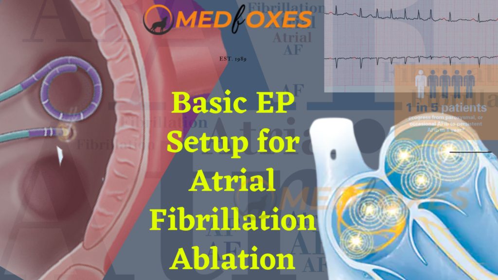
Catheter ablation for atrial fibrillation is becoming more common and is increasing in popularity around the world. Since pulmonary vein isolation has remained the foundation of atrial fibrillation ablation procedures, imaging and visualization technologies, as well as energy sources and energy delivery systems, have advanced significantly. To get good and safe results, you must have
Basic Setup for Atrial Fibrillation Ablation Read More »

Intracardiac Echocardiography catheters are designed for use in adult and adolescent pediatric patients to view cardiac architecture, blood flow, and other devices inside the heart. ICE has the potential to become one of the best ways to use imaging to guide interventions. Extremely aggressive techniques for electrophysiologic ablation therapy for the treatment of cardiac arrhythmias
Intracardiac Echocardiography Catheters Read More »
Non-Fluoroscopy ICE Catheter Manipulation is an evolving technique, Compared to transthoracic and transesophageal echo, ICE has a number of benefits. Higher resolution and picture quality are possible because of the substantially shorter image distances created by the probe’s intra-cardiac position. In contrast to the difficult transesophageal echo probe, it can also be done without anaesthesia.
Non-Fluoroscopy ICE Catheter Manipulation Read More »
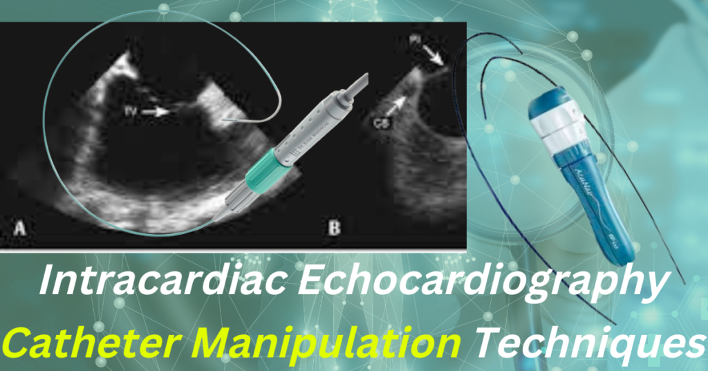
Intracardiac Echocardiography (ICE) enables direct visualisation of structures that are not normally visible with fluoroscopy or 3D anatomical mapping, monitoring for procedural problems, and real-time visualisation of catheters and cardiac structures. This article explains the techniques for acquiring views of all cardiac structures utilising a phased array ICE catheter. Catheter Manipulation Every ICE Console setup
Intracardiac Echocardiography Catheter Manipulation Technique Read More »