
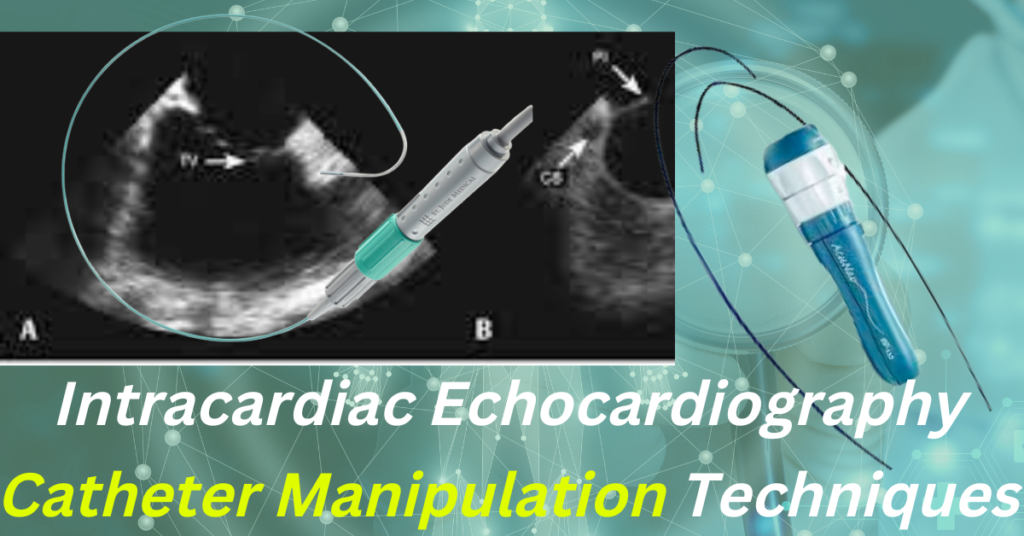
Intracardiac Echocardiography (ICE) enables direct visualisation of structures that are not normally visible with fluoroscopy or 3D anatomical mapping, monitoring for procedural problems, and real-time visualisation of catheters and cardiac structures. This article explains the techniques for acquiring views of all cardiac structures utilising a phased array ICE catheter.
Catheter Manipulation
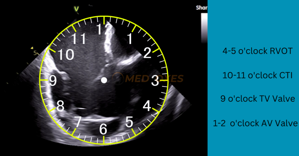
Every ICE Console setup can be completely customised. However, the default settings are left-to-right scrolling and an up-down setup, with the catheter’s tip at the top and in the middle of the display screen. A structure’s position on the main ICE screen is explained using a clock face analogy, with 12 o’clock being the top and centre of the screen and 6 o’clock being the bottom and centre of the screen.
Four fundamental techniques can be used to manipulate the catheter.
(1) Advancing/Withdrawing
(2) Anterior/Posterior Tilt
(3) Catheter rotation
(4) Right/Left Tilt.
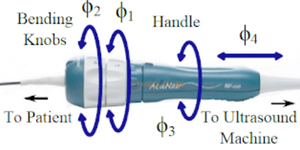
The resolution of the target structure can be improved greatly by even small movements in any of these directions.
To properly align the image in the centre of the view screen Advance the ICE catheter, which brings structures from the right side of the view screen (3 o’clock) to the centre of view. Withdrawing the catheter will bring structures from the left of the view screen (9 o’clock) to the centre of view.
Applying anterior tilt to the catheter will bring the tip of the catheter towards the structure in centre view, typically helping to bring that structure towards the 3 o’clock position.
Trying to apply posterior deflection will give the tip of the catheter away from the structure in centre view, typically bringing that structure towards the 9 o’clock position.
Clockwise (CW) rotation of the catheter itself, usually sweeps posteriorly, bringing posterior structures into centre view, for example, the coronary sinus os and left atrium (LA).
When rotating counterclockwise (CCW), which often sweeps forward, the aorta and RVOT come into view.
When entering cardiac chambers, it is very crucial to perform a small CW/CCW rotation to maintain a region of interest in focus.
The catheter tip will sweep and bend to the right in relation to the neutral position of the catheter when the right-to-left dial is turned to the right. The tip of that dial will bend and sweep to the left when it is turned to the left from neutral. This is especially helpful for fine-tuning the resolution of structures and evaluating the left atrial structures through the interatrial septum (IAS).
Catheter Positions to Obtain Images
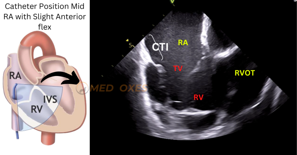
A) Home View : Move the ICE catheter slightly into the mid of the right atrium (RA) after the liver is seen on the screen. The appearing image on the screen is called Home Position and Visualizing RV, RA, and tricuspid valve (TV).
B) Right Atrium, Right Ventricular Outflow Tract (RVOT): Rotate the ICE catheter slightly counter-clockwise or to the patient’s left side, starting from the mid-right atrial home position with no steering applied. Just above the RVOT, the aortic valve will start to become visible.
C) Right Atrium, Left Ventricular Outflow Tract (LVOT): Rotate the catheter just a little bit clockwise while viewing it from the RVOT. In the centre of the screen, the left ventricular outflow tract (LVOT) and aortic valve will begin to appear. Just beyond the aortic valve, you might also be able to make out the pulmonary artery and/or pulmonary valve.
D) Right Atrium(RA), Coronary Sinus (CS), Left Atrium(LA), Mitral Valve(MV), Left Atrial Appendage(LAA): Rotate the ICE catheter clockwise from the RVOT view. In the same plane as the mitral valve, the coronary sinus (CS) will be seen directly below the right atrium. The left atrial appendage (LAA), also located in the same plane as the MV, will appear as an “out-pouching” from the left atrium (LA).