
Cardiac Magnetic Resonance Imaging is a three-dimensional technology that uses magnetic fields and radio waves to acquire detailed pictures of a patient’s heart.Unlike certain other imaging techniques, it doesn’t use ionising radiation, so patients don’t need to be exposed to harmful X-rays or radioactive substances.
In the assessment of aortic valve disease, cardiac MRI yields highly accurate information on the severity of aortic stenosis, as well as on procedural feasibility and device sizing. However, cardiac MRI has several contraindications like patients with metallic implants, pacemakers, or claustrophobia that limit its more widespread use in many cases.
New MRI indications on TAVI
As the number of TAVI procedures rises, similarly the number of patients who are unable to be examined properly using CTA, echocardiogram, or stress echocardiography will increase. As a result, non-contrast MR may be useful in the preoperative evaluation of the patients listed below.
Patients who have had a significant allergic reaction to intravenous iodinated contrast media and are unable to receive it for CTA.
Acute kidney injury or chronic kidney injury with serum creatinine > 2 mg/dL or GFR 30 mL/min/m2
Evaluating the severity of aortic stenosis in patients with poor acoustic windows and low cardiac outputs.
Using echocardiography to assess the severity of aortic stenosis in individuals who have moderate stenosis but are symptomatic for severe stenosis and are not candidates for stress echocardiography.
In the presence of significant delayed enhancement prior to TAVI, post-gadolinium delayed enhanced imaging has been utilised to determine the degree of coronary artery disease in patients undergoing TAVI, and it has been proven that there is a reduced LVEF recovery after TAVI. Gadolinium, on the other hand, should not be used in people who have weak renal function. Preoperative measures for TAVI do not require gadolinium enhanced MRI.
Aorta and Peripheral Arteries Evaluation
Although invasive angiography is the standard for evaluating the size and tortuosity of peripheral arteries, MR angiography may be a non-invasive alternative for patients with renal failure. A recent study indicates that gadolinium-enhanced MR angiography, using a new technique, can accurately assess iliac stenosis severity.[Fig. 1(A)][Fig. 1(B)].
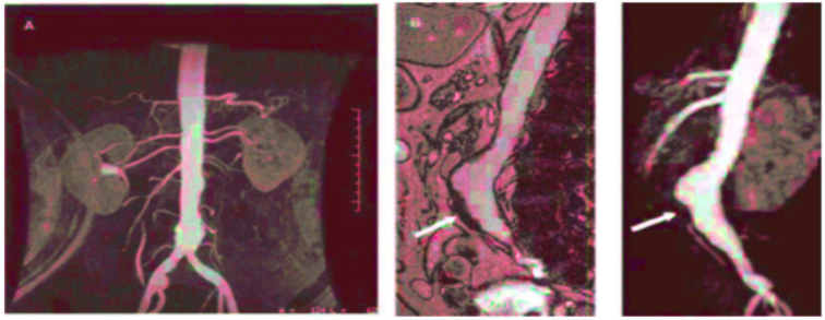
The diameter of the iliac arteries will determine whether a surgeon performs a transapical or a transfemoral approach. As well, calcifications within an abdominal aortic aneurysm can make the transfemoral approach difficult. However, MRI makes it more difficult to evaluate the extent and location of those calcifications.
Anatomy of the Aortic Valve and Valve Area Evaluation
The severity of aortic stenosis can be evaluated with magnetic resonance imaging, either by direct measurement of the stenotic orifice or by estimation of peak flow velocity. Several studies have demonstrated the accuracy of magnetic resonance imaging to estimate the aortic valve area. However, this measurement does not reflect the workload of the left ventricle, more related to the effective orifice area, usually smaller than the anatomical aortic valve area.
In this regard, velocity-encoded magnetic resonance imaging enables the assessment of the effective orifice area providing comparable assessments as those obtained with Doppler echocardiography.
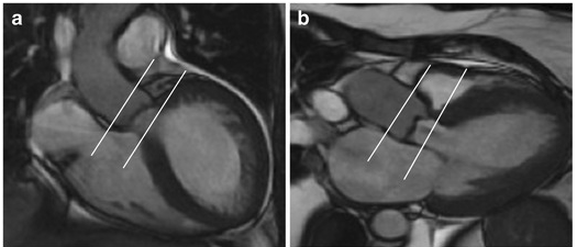
Aortic Annulus Sizing
The size of the aortic annulus is one of the cornerstones of procedural success. The ellipsoid geometry of the structure is bounded by a combination of muscular and fibrous components without a clear ring structure. Three-dimensional MRI and CT imaging clearly show that the annulus is smaller in the sagittal plane than in the coronal plane.
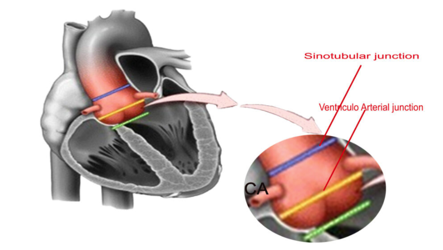
Evaluation of the Aortic Root and Coronary Artery
Measuring the sino-tubular junction is an important step in transcatheter aortic valve implantation since patients with a sino-tubular junction diameter ≥45mm are not good candidates for self-expandable devices. Before transcatheter aortic valve implantation, the coronary anatomy needs to be assessed. Although the feasibility of MRI for non-invasive angiography has been demonstrated, the accuracy of MRI to detect coronary artery stenoses is lower as compared to Multislice CT.
Further Reading:
Multislice CT Imaging on Transcatheter Aortic Valve Implantation
How Does Imaging Affect Transcatheter Aortic Valve Implantation?