
Contents
The nutritional supply of an embryo varies depending on its stage of development
- During the first week, before implantation, deutoplasm (accumulated cytoplasmic nutrients) of oocyte supplies nutrition.
- During the second week, breakdown products of endometrium (due to implantation) nourishes the embryo by simple diffusion.
- After the third week, maternal blood nourish embryo through utero placental circulation
The components of cardiovascular system can be studied as
Development of heart
Development of blood vessels
Constructing a Cardiogenic Zone
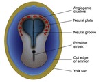
During the third week, cardiac progenitor cells develop just lateral to the primitive streak. These cells migrate through primitive streak cranially and forms horseshoe shaped primitive heart field in the splanchnopleuric mesoderm by end of the third week (Fig. 1)
On formation of head fold, primary heart field come to lie on dorsal side of pericardial sac. Endoderm of primitive pharynx induces vasculogenesis (formation of blood cells and vessels) in the primary heart field. Small vessels join to form two (right and left) endothelial heart tubes that give rise to the endocardium.
Splanchnopleuric mesoderm that lies between heart tube and pericardial cavity form a myoepicardial mantle. The myoepicardial mantle condense to form a) Myocardium (cardiac muscles) [cardiac muscles develop from splanchnopleuric mesoderm]. b) Epicardium (visceral layer of pericardium). The parietal layer of the pericardium is made up of somatopleuric mesoderm that surrounds the pericardial cavity.
Primitive heart starts beating on the 22nd day, By the 24th day, blood begins to circulate throughout the embryo.
Heart Tubes
Two heart tubes fuse together in the third week to form a single heart tube. The bifurcated ends of the heart tube remain. Its cranial end is known as the arterial end, whereas the caudal end is known as the venous end. From the cranial to the caudal end of the heart tube, five dilatations occur. 1. Truncus arteriosus 2. Bulbus cordis 3. Primitive ventricle 4. Primitive atrium 5. Sinus venosus (Fig. 2)
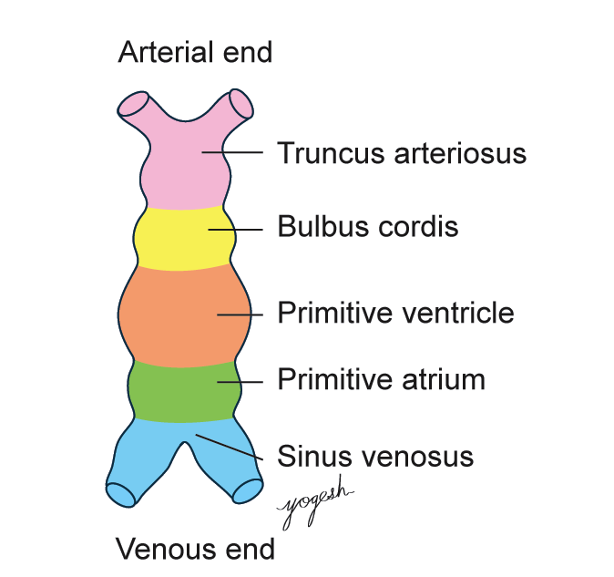
Parts of Heart Tube (Fig.2)
Ends of Heart Tube
Arterial end or truncus arteriosus shows right and left limbs. Through the first pair of pharyngeal arteries, these limbs (horns) are connected to the corresponding dorsal aorta. Soon six pairs of pharyngeal arch arteries connect truncus arteriosus with dorsal aorta. All pharyngeal arch arteries run on either side of foregut.
Venous End of Heart Tube
Unfused part of sinus venosus (venous end of heart tube) forms two horns (right and left). Each horn receives three veins (from lateral to medial) (Fig. 3) a) Common cardinal vein from the body wall b) Umbilical vein from the placenta c) Vitelline vein from the yolk sac.
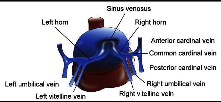
Fig 3
Derivatives of Heart Tube
Truncus arteriosus becomes Ascending aorta and Pulmonary trunk
Bulbus cordis becomes Conus arteriosus (smooth part of right ventricle) and Aortic vestibule (smooth part of left ventricle)
Primitive ventricle becomes Trabeculated part of right and left ventricles
Primitive atrium becomes Trabeculated part of right and left atrium
Sinus venosus becomes Right horn: Sinus venarum (smooth part of right atrium) and Left horn: Coronary sinus and oblique vein of atrium
ACQUISITION OF EXTERNAL FEATURES OF THE ADULT HEART
Heart tube undergoes growth and folding to acquire external features of the adult heart
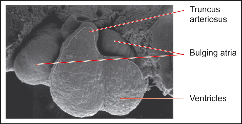
Fig 4
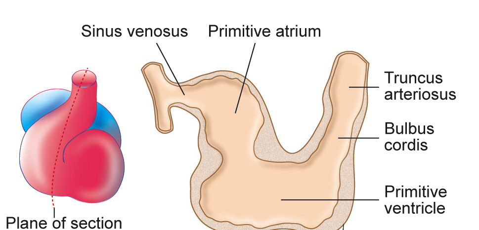
Fig 5
Initially, heart tube is placed longitudinally in pericardial cavity then Heart tube is suspended from dorsal wall of pericardial cavity by a fold of pericardium called dorsal mesocardium
Formation of bulboventricular loop: Bulbus cordis and primitive ventricle expand ventrally, forming a bulboventricular loop (U-shaped)
Formation of transverse sinus: The mesocardium connecting bulboventricular loop disappear to form a gap, that later called transverse sinus
Formation of S-loop: As primitive atrium and sinus venosus get freed from septum transversum, they come lie in the pericardial cavity dorso-cranial to the primitive ventricle and thus, S-shaped cardiac loop is formed.
Bulbus cordis and primitive ventricle are divided by bulboventricular sulcus, which eventually vanishes, and bulbus cordis and ventricle unite to form a single chamber.
Formation of auricles: Primitive atrium lies behind the truncus arteriosus. On expansion, primitive atrium project forward on either side of truncus arteriosus as auricles. (Fig.5)
Reference: Text book of Human embryology with clinical cases 8th edition , Moss and Adams 4th edition , braunwald cardiology, 6th edition and Healthnactive