MEDFOXES

Angiogram Radial Approach

Angiogram Radial Approach – The most compelling reason for adopting Trans-radial access is the increased patient safety that results from the virtual elimination of access site bleeding and vascular complications. In addition, TRA is associated with early sheath removal, improved patient comfort, faster recovery, and lower costs in comparison with transfemoral access Pre- procedure Assessment […]

Angiogram Radial Approach Read More »

Angiogram Femoral Approach

Angiogram, As of currently diagnostic coronary angiography remains the primary component of cardiac catheterization. The aim is to examine the whole coronary tree (both native vessels and any bypass grafts) while recording details of the coronary anatomy, including the following, the pattern of arterial distribution, anatomic or functional pathology (atherosclerosis, thrombosis, congenital anomalies, or focal coronary spasm),

Angiogram Femoral Approach Read More »

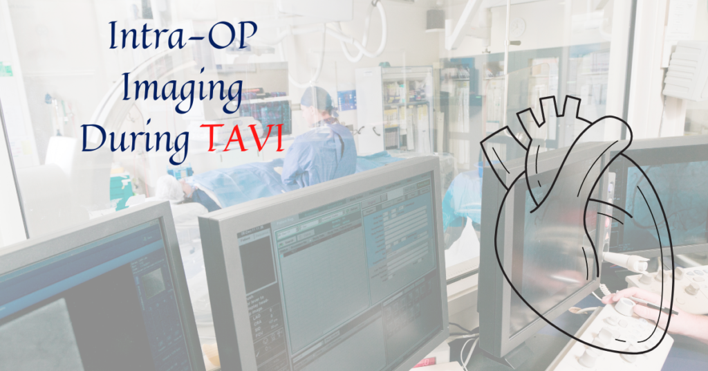
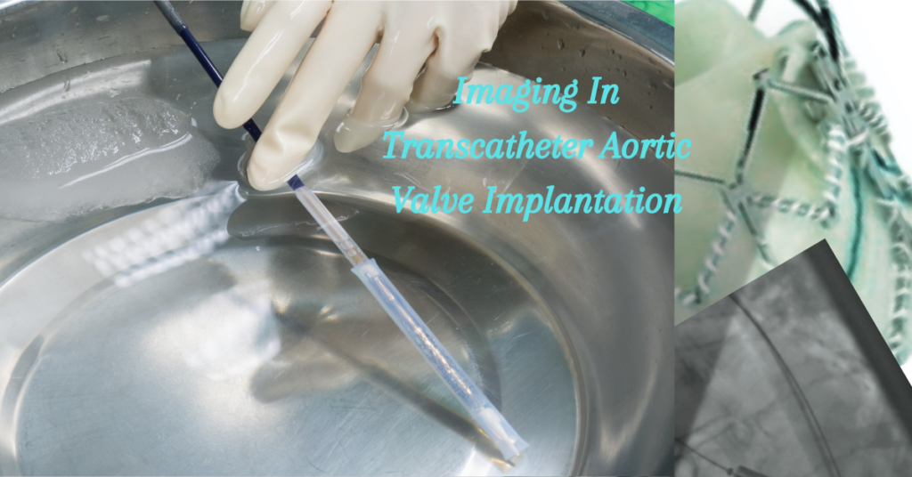
Intra-OP Imaging During TAVI

TAVI, just like any structural heart disease intervention, relies on effective imaging earlier, throughout, and after the procedure. Imaging’s role in percutaneous valve replacement is comparable to that of appropriate exposure and visualisation in open surgical valve replacement. Visualisation improves the speed, safety, and predictability of the procedure. In addition, imaging throughout the procedure is

Intra-OP Imaging During TAVI Read More »

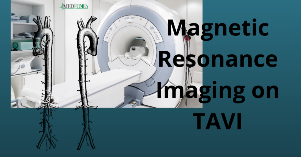
Magnetic Resonance Imaging on TAVI

Cardiac Magnetic Resonance Imaging is a three-dimensional technology that uses magnetic fields and radio waves to acquire detailed pictures of a patient’s heart.Unlike certain other imaging techniques, it doesn’t use ionising radiation, so patients don’t need to be exposed to harmful X-rays or radioactive substances. In the assessment of aortic valve disease, cardiac MRI yields

Magnetic Resonance Imaging on TAVI Read More »

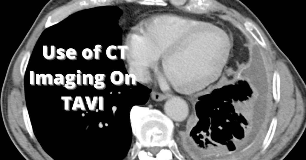
Multislice CT Imaging on Transcatheter Aortic Valve Implantation

Multislice computed tomography (MultiSlice CT) is the most comprehensive and non-invasive imaging modality to evaluate candidates for transcatheter aortic valve implantation. It can help confirm the severity of aortic stenosis and assess procedural feasibility, as well as device sizing. Evaluation of the Peripheral Arteries and Aorta Procedures to implant transcatheter aortic valves, particularly when done

Multislice CT Imaging on Transcatheter Aortic Valve Implantation Read More »

How Does Imaging Affect Transcatheter Aortic Valve Implantation?​

In the past few years, transcatheter aortic valve implantation has been one of the main breakthroughs in the treatment of patients with symptomatic, severe aortic stenosis who are at high risk for surgery. In patients with either self-expandable or balloon-expandable valvular prostheses, transcatheter aortic valve implantation has shown to be technically feasible and has provided

How Does Imaging Affect Transcatheter Aortic Valve Implantation?​ Read More »

Formation of Heart-Embryo changes

The nutritional supply of an embryo varies depending on its stage of development During the first week, before implantation, deutoplasm (accumulated cytoplasmic nutrients) of oocyte supplies nutrition. During the second week, breakdown products of endometrium (due to implantation) nourishes the embryo by simple diffusion. After the third week, maternal blood nourish embryo through utero placental

Formation of Heart-Embryo changes Read More »